ultrasound at 8 weeks
What to Expect During an Ultrasound at 8 Weeks
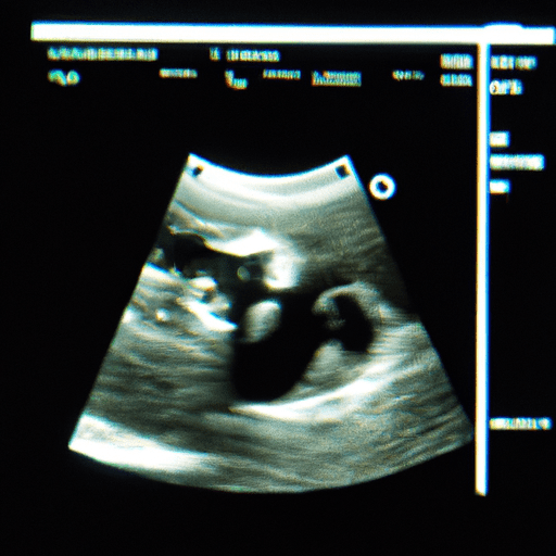
An ultrasound at 8 weeks is an important part of prenatal care. It is used to confirm the pregnancy, check the baby’s heartbeat, and assess the baby’s development. During the ultrasound, the technician will use a transducer to send sound waves through the uterus. These sound waves create an image of the baby on a monitor.
The technician will measure the baby’s size and check the baby’s heartbeat. They will also look for any signs of abnormalities. The technician may also measure the amount of amniotic fluid in the uterus. This helps to determine the baby’s health and development.
The ultrasound should take about 30 minutes. During the procedure, you may feel some pressure from the transducer. You may also feel some discomfort from the gel that is used to help the transducer move smoothly over your abdomen.
At the end of the ultrasound, the technician will provide you with a report of the findings. This report will include information about the baby’s size, heartbeat, and any abnormalities that were detected. Your doctor will review the report and discuss the results with you.
An ultrasound at 8 weeks is an important part of prenatal care. It helps to confirm the pregnancy, check the baby’s heartbeat, and assess the baby’s development. The procedure should take about 30 minutes and you may feel some pressure and discomfort from the transducer. At the end of the ultrasound, the technician will provide you with a report of the findings.
How Ultrasound Technology Has Improved Over Time
Ultrasound technology has come a long way since its inception in the 1950s. Initially used for medical imaging, ultrasound technology has since been applied to a variety of fields, including industrial, automotive, and aerospace engineering. Over the years, ultrasound technology has seen significant improvements in terms of accuracy, resolution, and cost-effectiveness.
One of the most significant improvements in ultrasound technology has been the development of higher-resolution imaging. This has allowed for more detailed images to be produced, which can be used to diagnose medical conditions more accurately. Additionally, the use of higher-resolution imaging has enabled the detection of smaller objects, such as tumors, which were previously undetectable.
Another major improvement in ultrasound technology has been the development of 3D imaging. This has allowed for more accurate diagnosis of medical conditions, as well as the ability to view the internal structure of organs and tissues. 3D imaging has also enabled the detection of smaller objects, such as tumors, which were previously undetectable.
The cost of ultrasound technology has also decreased significantly over the years. This has allowed for more widespread use of the technology, as it is now more affordable for medical professionals and patients alike. Additionally, the cost of ultrasound machines has decreased, making them more accessible to smaller medical facilities.
Finally, the accuracy of ultrasound technology has improved significantly over the years. This has allowed for more accurate diagnosis of medical conditions, as well as the ability to detect smaller objects, such as tumors, which were previously undetectable.
Overall, ultrasound technology has seen significant improvements over the years. These improvements have enabled more accurate diagnosis of medical conditions, as well as the ability to detect smaller objects, such as tumors, which were previously undetectable. Additionally, the cost of ultrasound technology has decreased significantly, making it more accessible to medical professionals and patients alike.
Common Ultrasound Findings at 8 Weeks
At 8 weeks of gestation, a standard ultrasound examination can provide a wealth of information about the developing fetus. Common findings include the presence of a fetal heartbeat, which is usually visible by this stage. The fetal heart rate is typically between 120 and 160 beats per minute. The size of the fetus can also be estimated, and is usually between 1.2 and 1.6 centimeters in length. The head and body are usually well-defined, and the arms and legs can be seen. The umbilical cord is usually visible, and the placenta can be seen in the uterus. The amniotic fluid surrounding the fetus can also be seen. In addition, the gender of the fetus may be visible, although this is not always the case.
The Benefits of Ultrasound at 8 Weeks
Ultrasound is a safe and non-invasive imaging technique used to visualize the internal organs and structures of the body. At 8 weeks, an ultrasound can provide a wealth of information about the developing fetus.
One of the primary benefits of an ultrasound at 8 weeks is the ability to confirm the gestational age of the fetus. This is important for determining the due date and monitoring the growth and development of the baby. The ultrasound can also detect the presence of multiple fetuses, which is important for planning the delivery and postnatal care.
The ultrasound can also detect the presence of any major structural abnormalities in the fetus. This includes the heart, brain, and other organs. It can also detect any major birth defects, such as spina bifida or anencephaly. This information can be used to plan for any necessary interventions or treatments that may be needed after birth.
The ultrasound can also detect the presence of any major chromosomal abnormalities, such as Down syndrome. This can be important for determining the risk of certain genetic disorders and planning for any necessary interventions or treatments that may be needed after birth.
Finally, the ultrasound can provide important information about the health of the placenta and umbilical cord. This can be important for monitoring the health of the fetus and ensuring that it is receiving adequate nutrition and oxygen.
Overall, an ultrasound at 8 weeks can provide a wealth of information about the developing fetus. This information can be used to plan for any necessary interventions or treatments that may be needed after birth. It can also provide important information about the health of the placenta and umbilical cord. For these reasons, an ultrasound at 8 weeks is an important part of prenatal care.
ultrasound at 8 weeks
How to Prepare for an Ultrasound at 8 Weeks
Preparing for an ultrasound at 8 weeks is an important step in ensuring a successful and accurate examination. An ultrasound is a non-invasive procedure that uses sound waves to create images of the inside of the body. It is used to diagnose and monitor a variety of medical conditions.
Before the ultrasound, it is important to make sure that you are well-prepared. Here are some tips to help you get ready for your 8-week ultrasound:
1. Schedule your appointment: Make sure to schedule your appointment in advance so that you have enough time to prepare.
2. Wear comfortable clothing: Wear loose-fitting clothing that is easy to remove. Avoid wearing any jewelry or metal objects that may interfere with the ultrasound.
3. Drink plenty of water: It is important to drink plenty of water before the ultrasound to ensure that the images are clear.
4. Empty your bladder: Make sure to empty your bladder before the ultrasound to ensure that the images are not distorted.
5. Bring a list of questions: Make sure to bring a list of questions that you may have for your doctor.
6. Bring a friend or family member: It is always a good idea to bring a friend or family member with you to the appointment.
By following these tips, you can ensure that you are well-prepared for your 8-week ultrasound. If you have any questions or concerns, make sure to speak to your doctor before the appointment.
What to Ask Your Doctor Before an Ultrasound at 8 Weeks
Before undergoing an ultrasound at 8 weeks, it is important to ask your doctor the following questions:
1. What type of ultrasound will be performed?
2. What is the purpose of the ultrasound?
3. How long will the procedure take?
4. What preparation is required prior to the ultrasound?
5. Will I be able to see the images of the ultrasound?
6. Are there any risks associated with the ultrasound?
7. What will the results of the ultrasound tell me?
8. Will I need to follow up with any additional tests or procedures?
By asking these questions, you can ensure that you are fully informed and prepared for your ultrasound at 8 weeks.
How to Interpret Ultrasound Results at 8 Weeks
Ultrasound results at 8 weeks of pregnancy can provide important information about the health of the fetus. During this stage of pregnancy, the ultrasound can be used to detect the presence of a heartbeat, the size of the fetus, and the number of fetuses present.
The presence of a heartbeat is an important indicator of the health of the fetus. During an ultrasound at 8 weeks, the technician will look for a heartbeat in the fetal pole, which is the area of the fetus that is visible on the ultrasound. If a heartbeat is detected, it is a good indication that the fetus is healthy and developing normally.
The size of the fetus is also important to monitor during this stage of pregnancy. The technician will measure the crown-rump length (CRL) of the fetus, which is the distance from the top of the head to the bottom of the buttocks. The CRL should be within a certain range for a fetus at 8 weeks.
If the CRL is outside of this range, it could indicate a potential problem with the development of the fetus.Finally, the technician will look for the presence of multiple fetuses. If more than one fetus is present, it is important to monitor the growth of each fetus to ensure that they are all developing normally.
Overall, an ultrasound at 8 weeks can provide important information about the health of the fetus. It is important to discuss the results of the ultrasound with a healthcare provider to ensure that the fetus is developing normally.
What to Expect During an Ultrasound at 5 Weeks