dentist x ray
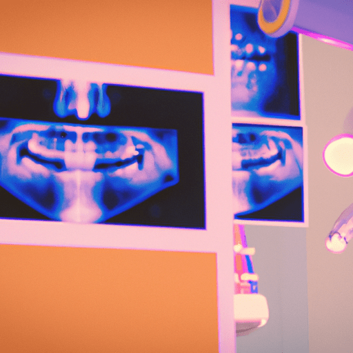
Digital X-rays are a revolutionary advancement in dental technology that has revolutionized the way dentists diagnose and treat their patients. Digital X-rays offer a number of benefits over traditional film X-rays, making them an invaluable tool for dentists.
The first major benefit of digital X-rays is that they are much faster and more efficient than traditional film X-rays. Digital X-rays can be taken in a matter of seconds, whereas traditional film X-rays can take up to several minutes. This means that dentists can take multiple X-rays in a single visit, allowing them to diagnose and treat their patients more quickly and efficiently.
Another major benefit of digital X-rays is that they produce much higher quality images than traditional film X-rays. Digital X-rays produce images that are much sharper and clearer than traditional film X-rays, allowing dentists to more accurately diagnose and treat their patients. Digital X-rays also allow dentists to zoom in on specific areas of the mouth, allowing them to get a better view of any potential problems.
Digital X-rays are also much safer than traditional film X-rays. Digital X-rays produce much lower levels of radiation than traditional film X-rays, making them much safer for both the patient and the dentist. This means that dentists can take multiple X-rays without worrying about exposing their patients to dangerous levels of radiation.
Finally, digital X-rays are much more cost-effective than traditional film X-rays. Digital X-rays require much less equipment and supplies than traditional film X-rays, making them much more affordable for dentists. This means that dentists can save money on X-ray costs, allowing them to pass those savings on to their patients.
Overall, digital X-rays offer a number of benefits over traditional film X-rays, making them an invaluable tool for dentists. Digital X-rays are faster, produce higher quality images, are much safer, and are more cost-effective than traditional film X-rays. For these reasons, digital X-rays are quickly becoming the standard in dental care.
in what food is magnesium
How to Prepare for a Dental X-Ray
Preparing for a dental x-ray is an important part of the process. It is important to follow the instructions of your dentist or dental hygienist to ensure the best results. Here are some tips to help you prepare for your dental x-ray.
1. Wear comfortable clothing: Wear clothing that is loose and comfortable. Avoid wearing any jewelry or metal objects that may interfere with the x-ray.
2. Remove any dental appliances: If you have any dental appliances such as dentures, retainers, or braces, make sure to remove them before the x-ray.
3. Follow the instructions of your dentist: Your dentist or dental hygienist will provide you with specific instructions on how to prepare for the x-ray. Make sure to follow these instructions carefully.
4. Avoid eating or drinking before the x-ray: Eating or drinking before the x-ray can interfere with the results. Make sure to avoid eating or drinking for at least an hour before the x-ray.
5. Relax: It is important to remain relaxed during the x-ray. Take deep breaths and try to stay as still as possible.
By following these tips, you can ensure that your dental x-ray is successful and that you get the best results.
The Latest Advances in Dental X-Ray Technology
Advances in dental x-ray technology have revolutionized the way dentists diagnose and treat oral health issues. The latest developments in this field have made it easier for dentists to detect and diagnose problems quickly and accurately.
Digital radiography is one of the most significant advances in dental x-ray technology. This technology uses digital sensors instead of traditional film to capture images of the teeth and surrounding structures. Digital radiography is faster, more accurate, and produces higher-quality images than traditional film radiography. It also reduces radiation exposure to the patient, making it a safer option.
Cone beam computed tomography (CBCT) is another important advancement in dental x-ray technology. This technology uses a cone-shaped x-ray beam to create 3D images of the teeth and surrounding structures. CBCT is especially useful for diagnosing complex dental problems, such as impacted teeth, root fractures, and cysts. It also provides detailed images of the jawbone, which can be used to plan dental implant placement.
Intraoral cameras are another important advancement in dental x-ray technology. These cameras are small, handheld devices that allow dentists to take high-resolution images of the teeth and surrounding structures. Intraoral cameras provide a more detailed view of the teeth and gums than traditional radiography, making it easier for dentists to diagnose and treat oral health issues.
These are just a few of the latest advances in dental x-ray technology. With these new technologies, dentists are better equipped to diagnose and treat oral health issues quickly and accurately.
The Different Types of X-Rays Used in Dentistry
X-rays are an important tool used in dentistry to diagnose and treat a variety of dental conditions. X-rays allow dentists to see inside the teeth and jaw, and to detect problems that may not be visible to the naked eye. There are several different types of X-rays used in dentistry, each of which has its own unique purpose.
The most common type of X-ray used in dentistry is the intraoral X-ray. This type of X-ray is taken inside the mouth and is used to detect cavities, bone loss, and other dental problems. Intraoral X-rays are also used to evaluate the position of teeth and to plan orthodontic treatment.
Another type of X-ray used in dentistry is the panoramic X-ray. This type of X-ray provides a wide view of the entire mouth, including the teeth, jaw, and sinuses. Panoramic X-rays are used to detect impacted teeth, cysts, tumors, and other abnormalities.
Cone beam computed tomography (CBCT) is a type of X-ray that produces 3-dimensional images of the teeth and jaw. CBCT is used to diagnose and plan treatment for complex dental problems, such as impacted teeth, root canal therapy, and dental implants.
Finally, bitewing X-rays are used to detect cavities between the teeth. Bitewing X-rays are taken by placing a small piece of film between the upper and lower teeth.
X-rays are an important tool used in dentistry to diagnose and treat a variety of dental conditions. Different types of X-rays are used for different purposes, and each type of X-ray provides valuable information that helps dentists provide the best possible care for their patients.
The Risks of Dental X-Rays
Dental X-rays are a common and important part of dental care. They provide valuable information about the health of your teeth and gums that cannot be obtained during a regular dental exam. However, like any medical procedure, there are risks associated with dental X-rays.
The most common risk associated with dental X-rays is radiation exposure. X-rays use a small amount of radiation to create an image of the teeth and surrounding structures. The amount of radiation used is very small and is considered safe. However, it is important to understand that any exposure to radiation carries some risk.
Another risk associated with dental X-rays is the potential for allergic reactions. Some people may be allergic to the chemicals used in the X-ray film or the chemicals used to develop the film. If you have a known allergy to any of these chemicals, it is important to let your dentist know before having an X-ray taken.
Finally, there is a risk of damage to the teeth or surrounding structures from the X-ray beam. This is rare, but it is possible for the X-ray beam to cause damage to the teeth or surrounding structures if the beam is not properly aimed. Your dentist will take steps to ensure that the X-ray beam is properly aimed and that the exposure time is kept to a minimum.
Overall, the risks associated with dental X-rays are very small. However, it is important to understand the risks and to discuss any concerns with your dentist before having an X-ray taken.
How to Interpret Dental X-Ray Results
Interpreting dental X-ray results can be a complex process. It is important to understand the basics of how to read and interpret X-ray images in order to make an accurate diagnosis.
The first step in interpreting dental X-ray results is to identify the structures that are visible on the X-ray. This includes the teeth, the jawbone, and any other structures that may be present. It is important to note any abnormalities or changes in the size or shape of the structures.
The next step is to look for any signs of disease or infection. This includes cavities, abscesses, cysts, tumors, and other abnormalities. It is important to note any changes in the size or shape of the teeth or jawbone, as well as any changes in the color of the X-ray.
Finally, it is important to look for any signs of trauma or injury. This includes fractures, dislocations, and other signs of trauma. It is also important to note any changes in the alignment of the teeth or jawbone.
By carefully examining the X-ray images and noting any changes or abnormalities, a dentist can make an accurate diagnosis and provide the appropriate treatment. It is important to remember that X-ray images are only one part of the diagnostic process and should be used in conjunction with other diagnostic tools such as physical examination and laboratory tests.
The Cost of Dental X-Rays and Insurance Coverage
Dental X-rays are an important part of preventive dental care. They provide valuable information about the health of your teeth and gums that cannot be obtained during a regular dental exam. The cost of dental X-rays can vary depending on the type of X-ray and the number of X-rays taken.
Most dental insurance plans cover the cost of X-rays as part of preventive care. Generally, insurance plans will cover two bitewing X-rays per year and one full mouth series of X-rays every three to five years. Bitewing X-rays are used to detect cavities between the teeth, while a full mouth series of X-rays is used to detect any problems with the roots of the teeth, the jawbone, and other structures in the mouth.
If you need additional X-rays beyond what is covered by your insurance plan, you may be responsible for the cost. The cost of X-rays can range from $20 to $250, depending on the type of X-ray and the number of X-rays taken.
If you are concerned about the cost of X-rays, talk to your dentist about payment options. Many dentists offer payment plans or discounts for cash payments. Additionally, some dental insurance plans offer discounts for preventive care, such as X-rays.
It is important to remember that dental X-rays are an important part of preventive dental care. Regular X-rays can help detect problems early, which can save you time, money, and discomfort in the long run.