What to Expect During an Ultrasound at 5 Weeks
An ultrasound at 5 weeks is an important part of prenatal care. It is used to confirm a pregnancy and to check the health of the fetus. During the ultrasound, a technician will use a transducer to send sound waves into the uterus. These sound waves create an image of the fetus on a monitor.
The technician will measure the size of the fetus and check the heartbeat. The heartbeat can usually be seen and heard at this stage. The technician will also look for any signs of abnormalities.
The ultrasound will take about 30 minutes. During the procedure, you may be asked to drink a glass of water to fill your bladder. This will help the technician get a better view of the fetus.
You may also be asked to change positions during the ultrasound. This will help the technician get a better view of the fetus.
At 5 weeks, the fetus is still very small. It may be difficult to see the fetus clearly. The technician may need to take several images to get a clear picture.
After the ultrasound, the technician will discuss the results with you. They will explain any abnormalities they may have seen and answer any questions you may have.
An ultrasound at 5 weeks is an important part of prenatal care. It is used to confirm a pregnancy and to check the health of the fetus. With the help of a trained technician, you can get a better understanding of your pregnancy and the health of your baby.
How to Prepare for an Ultrasound at 5 Weeks
Preparing for an ultrasound at five weeks can be an exciting experience. An ultrasound is a safe and painless procedure that uses sound waves to create images of the inside of your body. It is used to check the health of your baby and to confirm your pregnancy. Here are some tips to help you prepare for your ultrasound at five weeks.
1. Schedule your appointment: Make sure to schedule your appointment with your doctor or midwife as soon as possible. This will ensure that you have enough time to prepare for the ultrasound.
2. Wear comfortable clothing: Wear comfortable clothing that is easy to remove. You may need to take off your clothing from the waist down for the ultrasound.
3. Drink plenty of water: Drink plenty of water before your appointment. This will help to fill your bladder, which will make it easier for the technician to get a clear image of your baby.
4. Empty your bladder: Make sure to empty your bladder before the ultrasound. This will help to make the images clearer.
5. Bring a support person: Bring a support person with you to the appointment. This will help to make the experience more comfortable and enjoyable.
By following these tips, you can ensure that you are well-prepared for your ultrasound at five weeks. An ultrasound is a safe and painless procedure that can provide you with valuable information about your baby’s health.
What to Look for in an Ultrasound at 5 Weeks
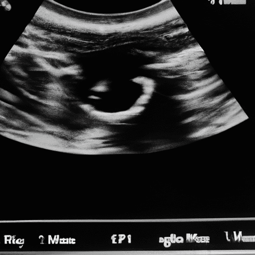
At five weeks pregnant, an ultrasound can provide a wealth of information about the health of the fetus. During the ultrasound, the technician will measure the size of the gestational sac, the yolk sac, and the embryo. The gestational sac is the fluid-filled sac that contains the embryo and is usually visible at five weeks. The yolk sac is a small round structure that provides nourishment to the embryo and is usually visible at five weeks. The embryo is the developing fetus and should be visible at five weeks.
The technician will also measure the crown-rump length (CRL) of the embryo. This is the length of the embryo from the top of its head to its bottom and is used to estimate the age of the fetus. The technician will also look for a heartbeat, which should be visible at five weeks.
The technician will also look for any signs of abnormalities. This includes any signs of a blighted ovum, which is when the gestational sac is present but the embryo is not. The technician will also look for any signs of a multiple pregnancy, such as two gestational sacs or two embryos.
Finally, the technician will look for any signs of ectopic pregnancy, which is when the embryo implants outside of the uterus. This can be a dangerous condition and should be monitored closely.
Overall, an ultrasound at five weeks can provide a wealth of information about the health of the fetus. It can help to detect any abnormalities and provide an estimate of the age of the fetus.
Common Ultrasound Findings at 5 Weeks
What to Expect During an Ultrasound at 5 Weeks
What to Expect During an Ultrasound at 5 Weeks
At five weeks gestation, a transabdominal ultrasound can be used to assess the development of the fetus. Common findings include a gestational sac, yolk sac, and fetal pole. The gestational sac is a fluid-filled structure that is seen as a white rim around a clear center. It is typically located in the uterus and is the first structure to be seen on ultrasound. The yolk sac is a small, round structure located within the gestational sac. It is seen as a white circle and is the source of nutrition for the developing fetus. The fetal pole is a thickening on the margin of the yolk sac and is the first visual evidence of the developing embryo. It is seen as a white line and is usually visible by five weeks gestation. Additionally, a heartbeat may be seen at this stage.
The Benefits of an Ultrasound at 5 Weeks
Ultrasounds are an important part of prenatal care, and at five weeks pregnant, an ultrasound can provide a wealth of information about the health of the fetus. An ultrasound at five weeks can help to confirm a viable pregnancy, as well as provide information about the location of the pregnancy, the number of fetuses, and the gestational age of the fetus.
An ultrasound at five weeks can help to confirm the presence of a heartbeat. This is an important indicator of a viable pregnancy, as a heartbeat is usually detectable at this stage. The ultrasound can also help to determine the location of the pregnancy. If the pregnancy is located in the uterus, it is considered a viable pregnancy. If the pregnancy is located outside of the uterus, it is considered an ectopic pregnancy and requires medical attention.
The ultrasound can also help to determine the number of fetuses present. This is important information for parents who are expecting multiples. The ultrasound can also help to determine the gestational age of the fetus. This is important information for parents who are trying to plan for the birth of their baby.
In addition to providing important information about the health of the fetus, an ultrasound at five weeks can also provide parents with a first glimpse of their baby. This can be an exciting moment for parents, as they get to see their baby for the first time.
Overall, an ultrasound at five weeks can provide a wealth of information about the health of the fetus and can be an exciting moment for parents. It is important to speak with your doctor to determine if an ultrasound is right for you.
How to Interpret an Ultrasound at 5 Weeks
At five weeks, an ultrasound can provide a wealth of information about the development of a fetus. It is important to note that the accuracy of the ultrasound can vary depending on the skill of the technician and the quality of the equipment.
The first thing to look for is the gestational sac. This is a fluid-filled sac that contains the embryo. It should be visible at five weeks. The gestational sac should be round and measure approximately two to three millimeters in diameter.
The next thing to look for is the yolk sac. This is a small, round sac that is located within the gestational sac. It should measure approximately one to two millimeters in diameter. The yolk sac is important because it provides nourishment to the developing embryo.
The third thing to look for is the fetal pole. This is the earliest visible sign of the developing embryo. It should measure approximately five millimeters in length. At this stage, it is not possible to determine the gender of the fetus.
Finally, it is important to look for a heartbeat. This should be visible at five weeks and should measure approximately 90 to 110 beats per minute.
By carefully examining the ultrasound images, it is possible to gain valuable insight into the development of the fetus at five weeks. It is important to remember that the accuracy of the ultrasound can vary depending on the skill of the technician and the quality of the equipment.
The Risks of an Ultrasound at 5 Weeks
Ultrasounds are a common procedure used to monitor the development of a fetus during pregnancy. An ultrasound at 5 weeks is generally considered safe, but there are some risks associated with the procedure.
The most common risk associated with an ultrasound at 5 weeks is the potential for inaccurate results. This is because the fetus is still very small and the ultrasound may not be able to detect certain abnormalities. Additionally, the accuracy of the results can be affected by the position of the fetus, the amount of amniotic fluid, and the quality of the ultrasound equipment.
Another risk associated with an ultrasound at 5 weeks is the potential for misdiagnosis. This is because the fetus is still developing and it can be difficult to accurately diagnose certain conditions. Additionally, the ultrasound technician may not be experienced enough to accurately interpret the results.
Finally, there is a risk of exposure to radiation during an ultrasound. While the amount of radiation used during an ultrasound is very low, it is still possible for the fetus to be exposed to a small amount of radiation.
This risk is generally considered to be very low, but it is still important to be aware of it. Overall, an ultrasound at 5 weeks is generally considered safe, but there are some risks associated with the procedure. It is important to discuss these risks with your doctor before undergoing an ultrasound.